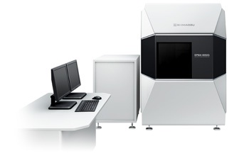
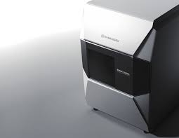
EPMA-8050G
Debut of the Grand EPMA

Optical System,
the Ultimate in Advanced
Shimadzu EPMA Analysis Performance
This instrument is equipped with a cutting-edge FE electron optical system, which provides unprecedented spatial resolution under all beam current conditions, from SEM observation conditions up to 1 μA order. Integration with high performance X-ray spectrometers that Shimadzu has fostered through the company’s traditions achieves the ultimate advance in analysis performance.
It is certainly appropriate to call this the grand EPMA, the debut of the ultimate EMPA system.
Electron Probe Microanalyzer
Ultra High Resolution Mapping
A mapping analysis of Sn balls on carbon was performed at a magnification of 30,000x. Even Sn particles a mere 50 nm in diameter, visible in the SE image (left), can be confirmed precisely in the X-ray image (right).

The Benefits of Cutting-Edge Technology
■ Unprecedented Spatial Resolution
The secondary-electron image resolution of 3 nm (30 kV accelerating voltage) is the highest level for an EPMA system.
This is the ultimate secondary-electron image resolution under analysis conditions.
(With an accelerating voltage of 10 kV, 20 nm at 10 nA / 50 nm at 100 nA / 150 nm at 1 μA)
Highest Secondary-Electron Image Resolution of 3 nm
This is a sample observation of gold particle deposition on carbon. A maximum resolution of 3 nm (at 30 kV) is achieved. The beam is focused even at a comparatively large beam current, so a smooth, high-resolution SEM image is easily obtained.

■ Large Beam Current Enabling Ultra High Sensitivity Analysis
This system achieves a maximum beam current of 3.0 μA (30 kV accelerating voltage).
There is no need to replace the objective aperture across the entire beam current range.
■ Up to Five High Performance 4-Inch X-Ray Spectrometers Can be Mounted
The 52.5° X-ray take-off angle is in a class by itself.
The 4-inch Rowland circle radius provides both high sensitivity and high resolution.
The system can accommodate up to five X-ray spectrometers of the same type.
■ Simple and Easy-to-Understand Operations for All Analyses
Advanced operability ensures that all operations can be performed with just a mouse.
The user interface is designed to be easy to understand.
Easy mode analysis automates all processes up to generating reports.
Ultra High Sensitivity Mapping
A mapping analysis of stainless steel was performed with a beam current of 1 μA at a magnification of 5,000x. (Left) The distribution of phases with slightly different Cr concentrations is precisely captured. (Right) The system succeeds in visualizing a distribution of Mn content under 0.1 %.

Advanced Technology Enabling Ultra High Sensitivity Analyses of Minute Areas

1. High-Brightness Schottky Emitter
A high output Schottky emitter with a larger tip diameter than ordinarily used in SEMs is adopted for the FE electron gun. A stable electron beam is obtained that while bright has the large current indispensable for high sensitivity analysis.
2. Special EPMA Electron Optical System
The electron optical system has a proprietary configuration and control method (Japan Patent No. 4595778). The condenser lens is set as close as possible to the electron gun. Crossover is formed not with the condenser lens but with an iris lens, with the objective aperture arranged at the same position. While this is a simple lens configuration, a large current can be obtained. At the same time, the angular aperture can be optimally configured under all current conditions, minimizing the electron beam diameter. Naturally, there is no need to replace the objective aperture.
3. Ultra High Vacuum Evacuation System
A two-stage differential evacuation system has been adopted, partitioned at the orifices between the electron gun chamber, intermediate chamber, and analysis chamber. Minimizing the orifice between the intermediate chamber and analysis chamber limits the flow of gas to the intermediate chamber. The electron gun chamber is always maintained at an ultra high vacuum level, stabilizing emitter operation.
4. X-Ray Spectrometers with High Sensitivity
The system can be equipped with up to five 4-inch X-ray spectrometers which provide both high sensitivity and high resolution.
The 52.5° X-ray take-off angle enhances the spatial resolution of the X-ray signal, while enabling high sensitivity analysis with minimal X-ray absorption by the sample.


